Super Miniature Fluorescence Microscopic Imaging System(TINISCOPE Series)
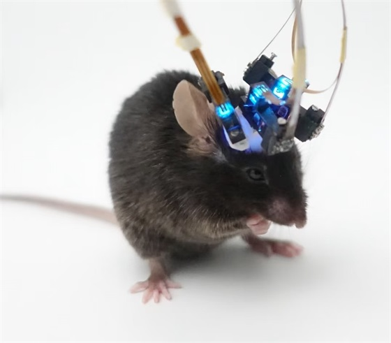
The Super Miniature Fluorescence Microscopic Imaging System (TINISCOPE Series) employs a smaller CMOS chip with serial output functionality, eliminating the need for a serializer chip. Additionally, it integrates the power support chip and oscillator functions into the Data Acquisition (DAQ) module. This innovative design significantly reduces the volume and weight of the CMOS module, enabling users to achieve synchronous imaging of essentially any four target brain regions. Tests have shown that mice can move freely without significant impact when wearing four devices simultaneously.
The TINISCOPE offers neuroscientists a brand-new and crucial research tool, facilitating the exploration of neuronal-level cross-brain region coordination mechanisms in animals related to perception, cognition, and behavior.
Technical Features
-
01
Ultra-Lightweight
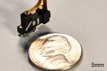
< 0.5 g,World's Lightest Head-Mounted Fluorescence Microscope
-
02
Ultra-Compact
Mice can wear four devices simultaneously, and their free movement is not significantly affected.
-
03
Wide-FOV imaging
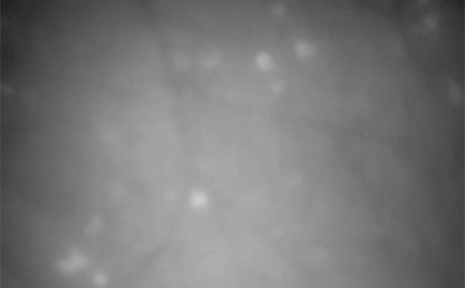
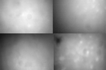
Under the premise of ensuring neuronal resolution, it is possible to collect signals from nearly a thousand neurons.
-
04
Ultra-long-term imaging
Manual or automatic unwinding solutions, Worry-free long-term imaging
-
05
High flexibility
The frame rate, exposure time, and gain of the microscope imaging are adjustable.
-
06
Efficient signal transmission
Synchronous transmission of four-channel calcium fluorescence signals and behavioral video signals
Application examples
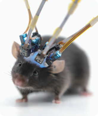
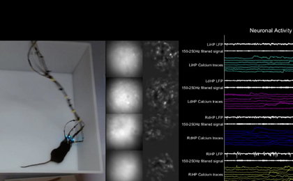
Mice equipped with four microscopes
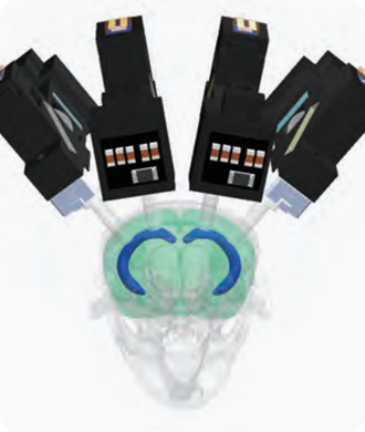
Microscope Imaging Site Model Diagram
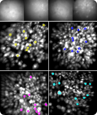
Quadruple-Brain-Region Simultaneous Imaging Results
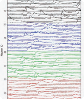
Population Neuronal Calcium Fluorescence Intensity Traces
Product Specifications
- Imaging ModeSingle-Photon Microscopy
- Excitation LightMiniaturized Integrated LED Light Source
- NA of the Objective Lens0.52
- Imaging Field of View720*540 μm
- Working Distance0 -250 μm
- Intracranial Focusing Depth0-250 μm
- Sampling Frequency25-40 Hz
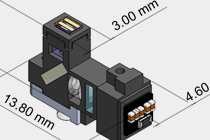
- Main Body Dimensions4.6 mm X 13.8 mm X 8.24 mm
- Weight<0.5 g
- Cable Length0.7 m -2 .7 m
- GRINIENS Diameter0.3 mm-1mm
- External Trigger InterfaceBNC X 4
- Data TransmissionUSB3.0 X 2 or USB3.0 X 4
- Magnification2.67x
- Pixel Resolution1μm
- Behavioral CameraSupport for synchronous acquisition
- Excitation Spectrum470±20 nm
- Excitation Intensity Range0 - 2.5 mW/mm²
- Emission Spectrum525 ± 25 nm
- Imaging TypeGrayscale
- ADC Precision10 bit
- Inter-lens Distance<1mm