High-Speed 3D Fluorescence Microscope for Large Samples
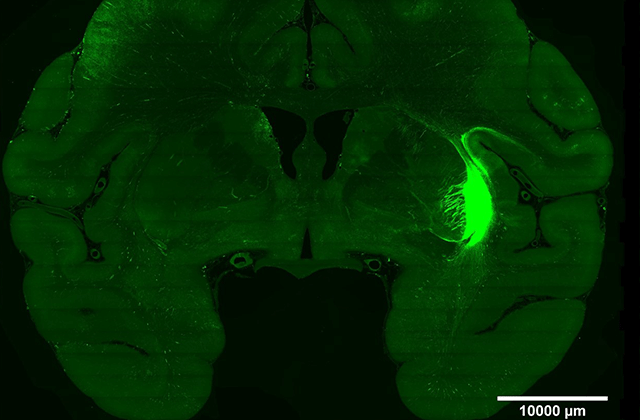
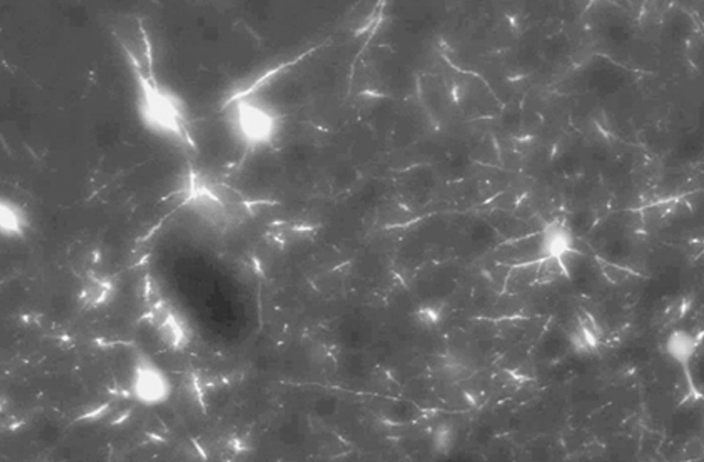
Based on proprietary Volumetric Imaging with Synchronous on-the-fly-scanning and Readout (VISoR) technology, our High-Speed Fluorescence Microimaging System can complete imaging of a whole mouse brain sample within 2 hours at a voxel resolution of 0.5x0.5x3.5μm³ (or 0.5 hour 1x1x2.5μm³), achieving efficient mesoscopic mapping of the entire brains from mouse to monkey.This system enables continuous 3D imaging of thick tissue samples, significantly enhancing data acquisition rates. By integrating thick tissue section clearing techniques and 3D data reconstruction technologies, the system is suitable for multi-channel imaging of samples of various sizes, including mouse brains, tree shrew brains, marmoset brains, macaque brains and even human brains. Compared to confocal microscopes and two-photon fluorescence microscopes, VISoR technology offers imaging speeds that are hundreds to thousands of times faster, making it an essential tool for analyzing brain structure and function. lt also has extensive applications in histology and medical fields.

Our Technology
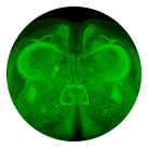
VISoR imaging technology is a novel rapid 3D micro-resolution imaging system developed by Professor Guoqiang Bi's research group at the University of Science and Technology of China (Wang et al., 2019, Nat Sci Rev). By integrating brain tissue clearing techniques and 3D reconstruction technologies, VISoR can achieve synaptic-level resolution of an entire mouse brain within 1.5 hours at a voxel resolution of 0.5μm x 0.5μm x 3.5μm. This technology enables continuous 3D imaging of thick tissue samples, significantly enhancing data acquisition rates. It combines thick tissue section clearing techniques with 3D data reconstruction technologies, making it suitable for multi-channel imaging of samples of various sizes, including mouse brains. Compared to confocal microscopy and two-photon fluorescence microscopy, VISoR offers a 10 to 100-fold increase in data acquisition rates. Both its imaging speed and resolution surpass those of traditional slice-scanning microscopes. VISoR can be used for rapid analysis in experiments such as viral tracing and immediate early gene expression analysis, providing fast insights into behavior-related neural circuits. Its capabilities make it an essential tool for quickly analyzing complex biological systems and advancing our understanding of brain structure and function.
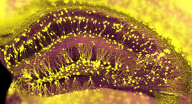
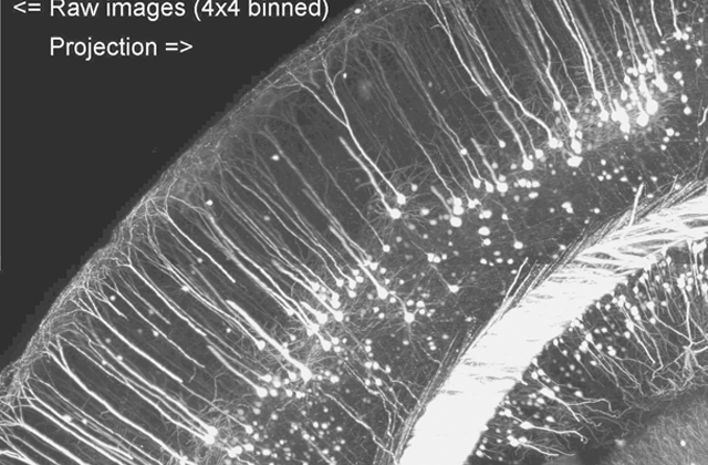
Whole-brain image reconstruction using VISoR technology can be completed with data acquisition in just 1.5 hours at a resolution of 0.5μm x 0.5μm x 3.5μm, generating approximately 3TB of data.
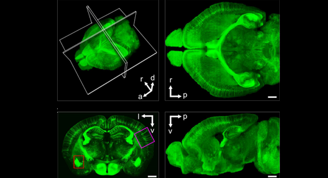
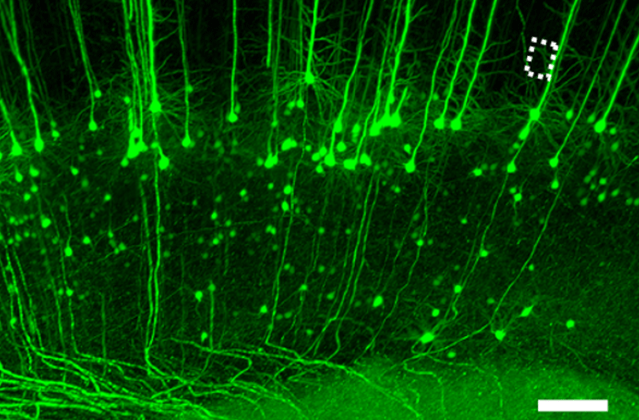
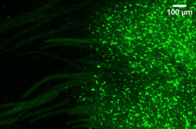
1.5 hours to complete synaptic-resolution whole-brain imaging of THY1-YFP mice
Imaging Figure
3D Rapid Pathological Diagnosis
More Information
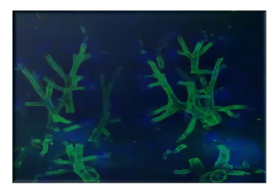
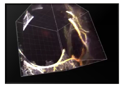
3D diagnostics provide more information compared to 2D, allowing for the capture of the entire sample's full information, resulting in more accurate diagnostic outcomes.
Certain specific diseases can only be diagnosed using three-dimensional diagnostics.
For example, the nature of oral mucosal cancer must be determined based on its growth along the Z-axis.
Faster speed
It improves detection efficiency by over a hundredfold compared to traditional methods and can be used for intraoperative detection
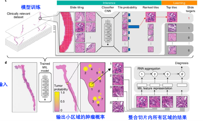
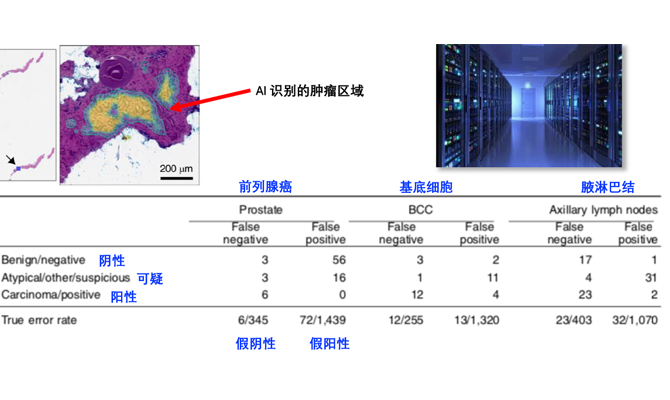
AI Pathology Analysis
Digital Human
Digital Human is the latest frontier in medical technology, representing a highly interdisciplinary integration of medicine, life sciences, information science, intelligent science and technology, and computer science. Research in this field holds significant implications for medicine and related disciplines, and it is poised to profoundly transform the future.


